
Figure 6.
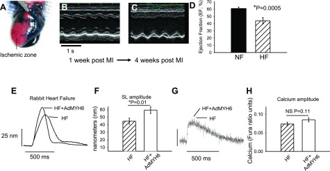
α-MyHC gene transfer increases the contractility of failing rabbit cardiac myocytes. A) An ischemic model of heart failure was implemented in adult rabbits. Successful coronary occlusion was confirmed by retrograde perfusion of Evans Blue dye. B, C) Representative m-mode echocardiography images of animals with a nonfailing heart (NF; B) and heart failure (HF; C) show that cardiac function was severely depressed after chronic LCX occlusion. D) Ventricular ejection fraction was depressed in HF vs. NF animals (43.9±2.2%, n=4 vs. 60.6±1.2%, n=4). E) Representative sarcomere length (SL) shortening measurements in HF cardiac myocytes 2 d after in vitro gene transfer with AdMYH6 (HF+AdMYH6) or with no virus treatment (HF). F) Summary data show that α-MyHC gene transfer increased SL shortening amplitude in electrically paced (1 Hz) single myocytes. HF amplitude = 44.6 ± 4.0 nm, n = 59; HF + AdMYH6 amplitude = 59.2 ± 4.5 nm, n = 50. G) Representative calcium transient recordings show no effect of AdMYH6 gene transfer on intracellular calcium cycling. H) Summary data show that the intracellular calcium transient amplitude was not significantly affected by α-MyHC gene transfer. HF amplitude = 0.074 ± 0.004, n = 53; HF + AdMYH6 amplitude = 0.085 ± 0.005 ratio units, n = 48.