
Figure 1.
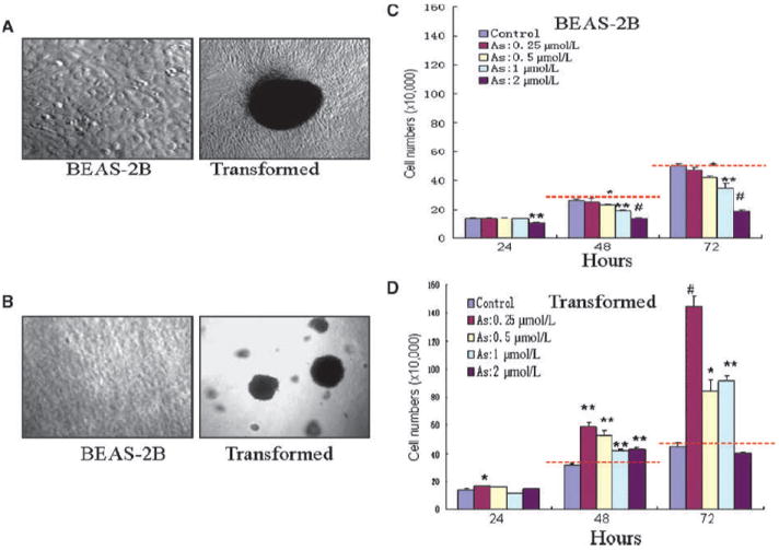
Continuous low concentration of As3+ exposure induces cell transformation. A, BEAS-2B cells maintained in As3+-free medium (left) or in a medium containing 0.25 to 2 μmol/L As3+ for 24 wk. Note the morphologic changes and multiple foci formation of the As3+-treated cells. B, As3+-transformed cells have features of anchorage-independent growth as determined by the formation of large colonies in soft agar. C, As3+ inhibits cell growth of the nontransformed cells (BEAS-2B) in a time- and dose-dependent manner. D, As3+ enhances cell growth of the transformed cells. For the purpose of comparison, red dash lines were drawn to indicate that the number of cells treated with As3+ was smaller than the control (C) or larger than the control. Columns, mean (n = 3); bars, SD. *, P < 0.05; **, P < 0.01; #, P < 0.001.