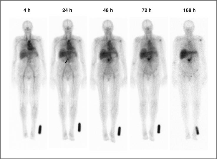
Figure 1.
Serial anterior whole-body planar scintigraphic images showing normal antibody biodistribution and tumor targeting in a patient with a recurrent mass in the pancreatic bed after undergoing a Whipple procedure. CT imaging had revealed a 1.5 × 2.1-cm mass surrounding the distal superior mesenteric artery. The images were acquired sequentially over 1 week, beginning 4 hours after administration of 111In-hPAM4. Image intensities were normalized by using an 111In standard near the left foot to correct for 111In physical decay and different acquisition times over this period. Uptake at the pancreatic site is clearly seen at 24 hours (arrow), and becomes progressively more prominent on subsequent images. Mild decreased intensity across the upper aspect of the liver is due to attenuation from breast tissue. Mild focal uptake in the left shoulder and pelvic area is of uncertain significance.

