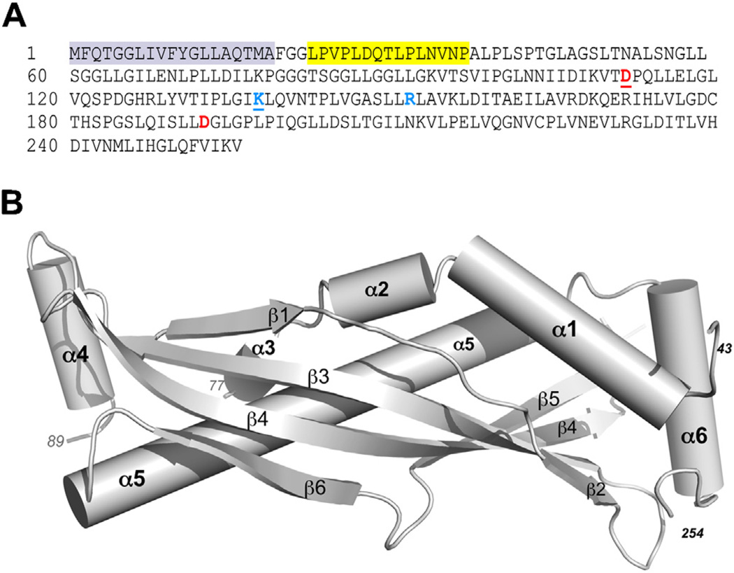
Fig. 1.
SPLUNC1 amino acid sequence and crystal structure. (A) The cleaved leader sequence is highlighted gray. The ENaC inhibitory domain (a.k.a. the S18 region) is highlighted in yellow. Basic (blue) and acidic (red) residues known to confer pH-sensitivity on SPLUNC1 are shown in bold. The underlined pair of residues constitute one potential salt bridge while the second set of residues constitute a second salt bridge. (B) Ribbon diagram of the crystal structure of human SPLUNC1.

