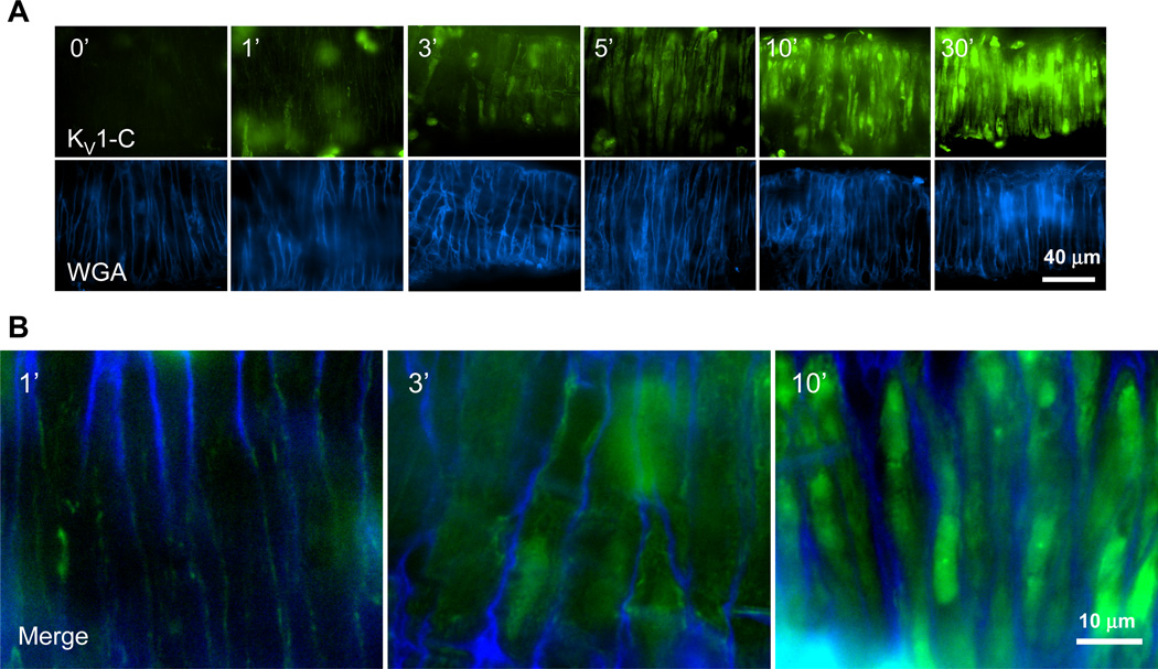
Figure 2. KV1-C peptide rapidly penetrates cVSMCs in CA.
A) Confocal images of isolated rat CA incubated with fluorescein-labeled KV1-C peptide (top row) for 0, 1, 3, 5, 10, and 30 min at 37°C. Alexa350-labeled wheat germ agglutinin (bottom row, WGA) was used as a cell surface marker for cVSMCs. Individual cVSMCs are visible vertically wrapping around the CA circumferentially, since the artery was placed horizontally for imaging. The brightness settings for the green channel in 10- and 30-minute treatments were reduced in order to display individual cells. Representative images from three similar experiments. B) Merged images from 1, 3, and 10-minute time points.

