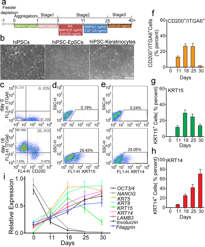
Figure 2. Staged differentiation of hiPSCs into human EpSCs.
a. An outline of the protocol used to differentiate hiPSCs to EpSCs and then mature keratinocytes. b. Morphologies of hiPSCs, hiPSC-derived EpSCs (hiPSC-EpSCs, obtained at day 18 after differentiation) and hiPSC-derived mature keratinocytes (hiPSC-keratinocytes, obtained at day 45 after differentiation). Scale bar, 100 µm. c-e. Flow cytometric analysis of CD200+/ITGA6+, KRT15+ and KRT14+ cells at day 0 and 18 during the differentiation. f-h. Quantitation of CD200+/ITGA6+, KRT15+ and KRT14+ cells by flow cytometric analysis. Data shown are mean ± SD of cell percentage from three independent experiments. i. qPCR analysis of OCT3/4, NANOG, KRT5, KRT8, KRT14, KRT15, LamB3, involucrin and filaggrin expression in hiPSC-derived cells at different stages of differentiation. Samples collected at day 0, day 11, day 18, day 25 and day 30 after differentiation were used for qRT-PCR analysis. Data shown are mean ± SD of the expression from three independent experiments.

