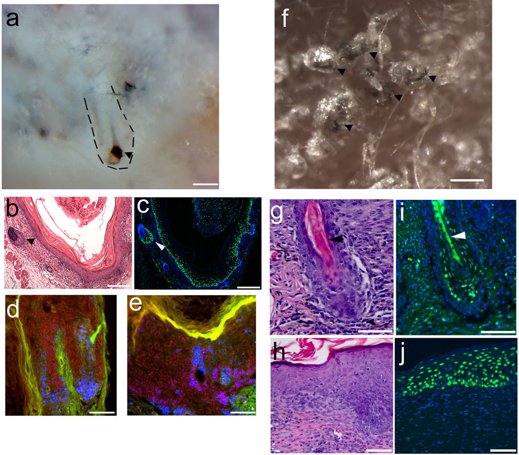
Figure 5. Folliculogeneic capacity of EpSCs derived from hiPSC tested in two different types of reconstitution assays.
a. hiPSC-derived CD200+/ITGA6+/SSEA3− cells form hair follicles in a patch reconstitution assay. hiPSC-derived CD200+/ITGA6+/SSEA3− cells were combined with mouse neonatal dermal cells and injected into the dermis of an immunodeficient mouse. After 3 weeks, hair follicles and hair follicle like structures were observed at the site of injection photographed from the underside of the skin. Dotted short lines outline a hair follicle. An arrowhead points to the pigmented bulb region of a hair follicle. Representative image from seven independent experiments. Scale bar, 500 µm. b. H&E staining of an epidermal cyst with attached hair follicles formed from hiPSC-EpSCs. An arrowhead points to a hair follicle. Scale bar, 200 µm. c. Human specific Alu probe staining (green nuclei) confirms human origin of follicular epithelium and epidermal cyst lining which were generated by hiPSC-derived EpSCs. An arrowhead points to a hair follicle. Scale bar, 200 µm. d, e. In situ hybridization using pan-centromeric probes specific for human (red) and mouse (green) respectively show human origin of follicular epithelium (d) and epidermal lining (e). Scale bar, 30 µm. f. Hair follicles form from hiPSC-derived CD200+/ITGA6+/SSEA3− cells and mixed with mouse neonatal dermal cells in a reconstitution assay using a silicone chamber. hiPSC-EpSCs and mouse neonatal dermal fibroblasts were mixed together and placed in a chamber transplanted onto the back skin of a nude mouse29. After three weeks, skin and hair follicles formed in the chamber. Pigmented human-like hair shafts were thicker than the surrounding mouse hair shafts. Arrowheads point to the pigmented hair shafts. Scale bar, 1 mm. g. H&E staining of a reconstituted hair follicle (H&E stain). An arrowhead points to the hair shaft. Scale bar, 150 um. h. A human-like multilayered epidermis was formed (H&E stain). Scale bar, 100 um. i,j. Human specific Alu probe staining of human hair follicles (i) and epidermis (j). Human cells were stained by human specific Alu probe labeled with FITC. An arrowhead points to the hair shaft (i). Scale bar, 150 um (i), 100 um (j). Representative images from three independent experiments.

