Figure 2.
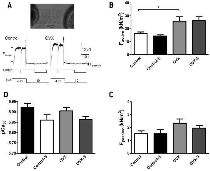
Effects of ovariectomy and stress on cardiomyocyte mechanics. (A) Single cardiomyocyte, isolated from rat myocardium, mounted between a sensitive force transducer and an electromagnetic motor (upper panel). Measurements of maximum (pCa [ie, -10log[Ca2+]] 4.75) Ca2+-dependent active (Factive) and Ca2+-independent (pCa 10) passive (Fpassive) force levels in control and ovariectomized (OVX) animals (lower panel). (B) Effect of ovariectomy (OVX) and stress (control-S and OVX-S) on cardiomyocyte Factive (*P < 0.050 vs control). (C) Calcium sensitivity of force production (pCa50) determined in skinned cardiomyocytes derived from LV tissue in the four animal groups. (D) Unaltered Fpassive by ovariectomy or stress (number of cardiomyocytes, n = 10 per group of 5-7 animals).

