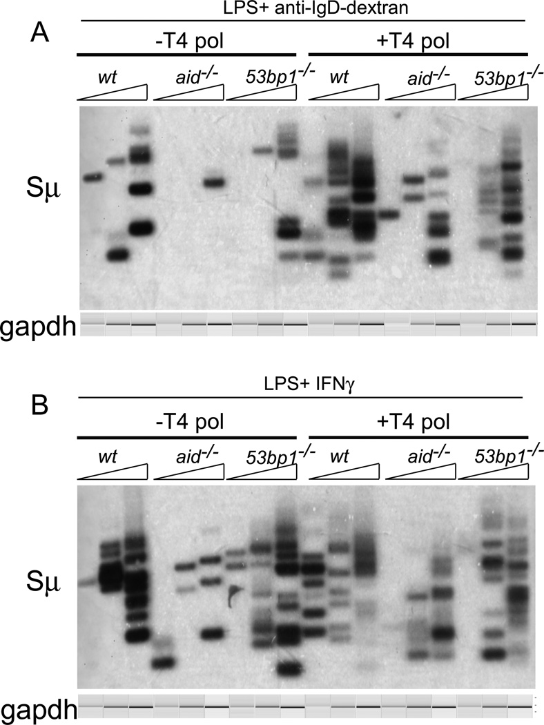
FIGURE 3. Both blunt and staggered Sµ DSBs are unaffected by the absence of 53BP1.
Sµ LM-PCRs were performed on 3-fold dilutions of DNA isolated from splenic B cells that had been stimulated to switch to IgG3 (A) or to IgG2a (B). As indicated, DNA was untreated or treated with T4 polymerase prior to linker ligation to identify staggered DSBs. Gels shown are from two independent experiments. The mb-1 PCR bands shown were obtained by electrophoretic analysis on a QIAxcel Advanced instrument, which subjects each sample to electrophoresis in a capillary, and provides an image and quantitation of each lane.

