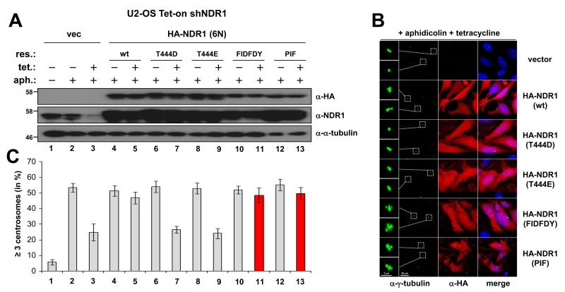
Figure 3. Active NDR1 versions drive centrosome overduplication, while NDR1(T444D) and NDR1(T444E) do not support centrosome amplification.
(A) U2-OS cells stably expressing tetracycline-regulated short-hairpin RNA (shRNA) directed against human NDR1 were infected with empty vector (lanes 1 to 3), HA-NDR1 wild-type (lanes 4 and 5), or hydrophobic motif mutants (lanes 6 to 13) that are refractory to shRNA [HA-NDR1 (6N)]. After incubation for 72 hours without (-) or with (+) tetracycline (2 μg/ml) and for a further 72 hours with aphidicolin (2 μg/ml), cells were analysed by immunoblotting with indicated antibodies. Molecular weights are shown. (B) In parallel, cells were processed for immunofluorescence with anti-γ-tubulin (green) and anti-HA (red) antibodies. DNA is stained blue. Insets show enlargements of centrosomes in green. (C) Histograms showing percentages of cells with excess centrosomes (≥ 3). Cumulative data from three independent experiments with at least two replicates of 100 cells counted per experiment. Error bars indicate standard deviations.

