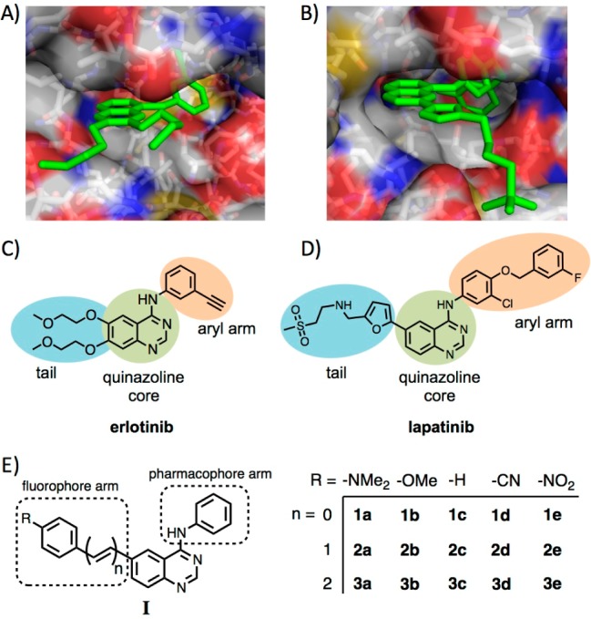
Figure 1.

Crystal structures of the EGFR ATP-binding pocket with (A) erlotinib (PDB ID: 1M17)15 and (B) lapatinib (PDB ID: 1XKK)9 reveal the inhibitor binding modes. The arms at the 6-position of the quinazoline core (in blue; C, D) may be replaced by fluorophore arms without disturbing the key binding contacts. (E) General structure and substituent key for the synthesized fluorescent quinazolines.
