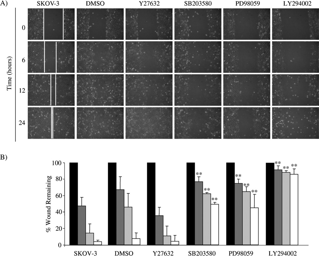
Fig. 1.
Rho kinase/ROCK, p38 MAPK, MEK and PI3K inhibitors differentially alter wound-induced SKOV-3 cell migration. (A) A wound-induced migration assay was performed on SKOV-3 cells in the absence and presence of DMSO vehicle control, 10 µM Y27632, 10 µM SB203580, 25 µM PD98059 or 25 µM LY294002. Photomicrographs of the treated cells are shown for the initial wounding (0 h) and at 6, 12 and 24 h post-wounding. (B) The distance the cells migrated into the wounded area was quantified as described in Materials and methods and presented as the percent wound remaining at the given times. **p<0.01, compared with untreated SKOV-3 cells at the given time point, 0 h (black bar), 6 h (dark grey bar), 12 h (light grey bar) and 24 h (white bar).

