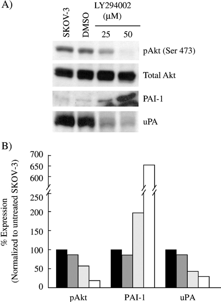
Fig. 3.
Inhibition of PI3K increases PAI-1 expression and decreases uPA expression in SKOV-3 cells. SKOV-3 cells were treated for 24 h in serum-free media with two doses of the PI3K inhibitor, LY294002. (A) Lysates were harvested and equal protein was separated on a 10% polyacrylamide gel, transferred and blotted for active Akt (phospho-Ser473) and total Akt. Conditioned media from the cells were concentrated and treated the same as the lysates, except blotted for PAI-1 and uPA, loaded by total protein. (B) The bands detected byWestern blot were quantitated by densitometry as described in Materials and methods as SKOV-3 cells untreated (black bar), DMSO-treated control SKOV-3 cells (dark grey bar), 25 µM LY294002 (light grey bar) and 50 µM LY294002 (white bar).

