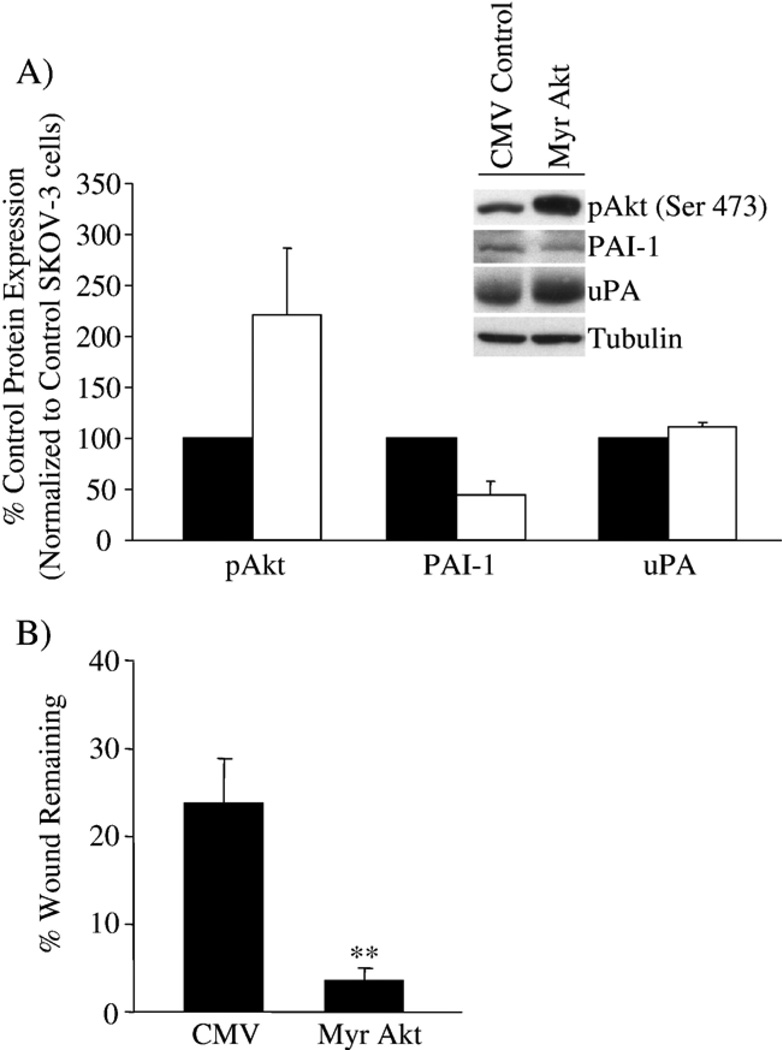
Fig. 7.
Over-expression of Akt in SKOV-3 cells affects PAI-1 expression and cell migration. SKOV-3 cells were infected with control CMV or Myr Akt adenovirus at MOI 50. (A) Active Akt (pAkt), PAI-1 and uPA expression were monitored, and densitometry used to quantify the change in protein expression detected by Western blot, normalized to tubulin as a protein loading control; the results are presented as the percent expression compared to control CMV adenovirus-treated SKOV-3 cells. Results are presented as the percent expression of Myr Akt-infected SKOV-3 cells (white bar) compared to CMV control adenovirus-infected SKOV-3 cells (black bar). (B) The effect of Myr Akt expression on SKOV-3 cell migration was measured in a wound-induced migration assay in 1% FBS conditions, and the results are presented as the percent wound remaining at 12 h. **p<0.01 compared with SKOV-3 cells with control CMV alone.

