
Figure 3. YAP KO mice have thin arterial walls and dilated arteries.
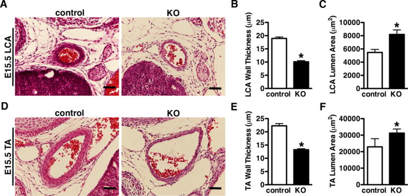
A. HE stained images of the left carotid artery (LCA) or thoracic artery (TA) (D) from E15.5 control and YAP KO embryos. The thickness of medium wall and size of lumen area were quantified and plotted as depicted in panel “B”, “C”, and “E”, “F”, respectively. Data were collected from 3 control and 5 KO embryos. *p<0.05. Scale bar: 50um.