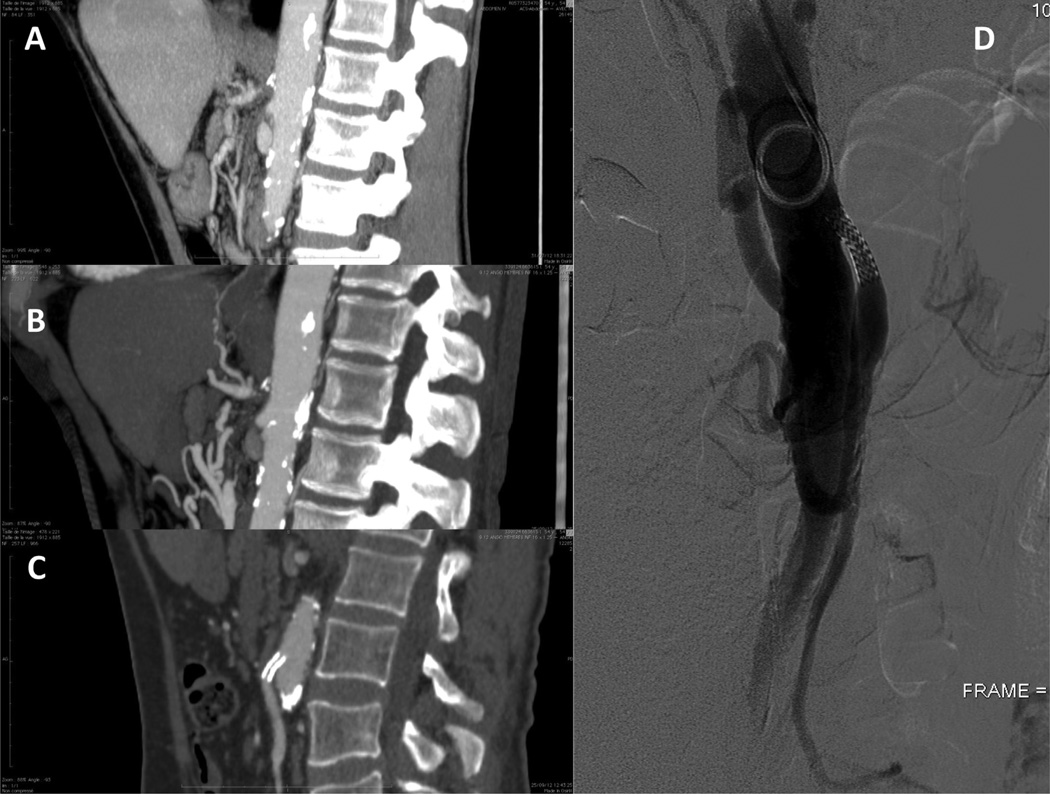
Figure 2.
Patient number 2. A computed tomography scan showed a 4.5-cm segment of thrombosed superior mesenteric artery, and stenosis of the celiac trunk, consistent with chronic mesenteric ischemia (A). At the end of the case, angiography showed perfusion of the Riolan arcade and branches of the inferior mesenteric artery (B). Postoperative computed tomography scan and 6-month follow-up angiogram showing patent stent in the inferior mesenteric artery (C, D).

