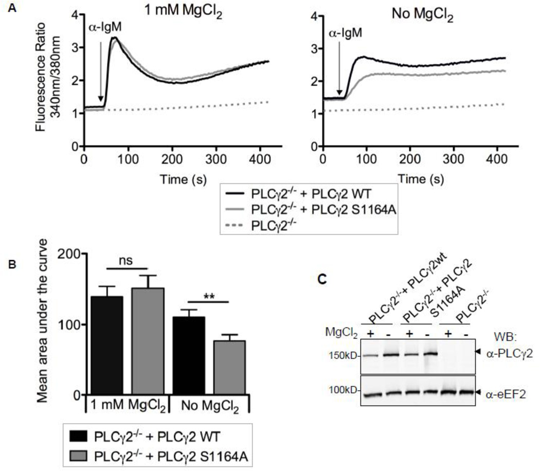
FIGURE 4. Under hypomagnesic conditions BCR mediated Ca2+-responses are reduced in PLCγ2−/− DT40 cells complemented with PLCγ2-S1164A.
A, Changes in free cytosolic Ca2+ in PLCγ2−/− cells alone or with stable expression of PLCγ2 WT or PLCγ2 S1164A. Cell lines were cultured in 0 or 1 mM MgCl2 for 15–25 h and loaded with Fura-2 for 30 min. Cells were stimulated with 1.2 µg anti-chicken IgM and analyzed by a fluorometer. Shown traces are representative of 4 separate experiments. B, Quantification of the results presented in A and B where bars represent the mean area under the curve and a paired two-tailed t-test was performed, mean ± SEM; **p < 0.01. C, Immunoblot of 2.5 × 10⁵ equivalent cells from whole cell lysates made from A and probed with anti-PLCγ2 and anti-eEF2 (loading control).

