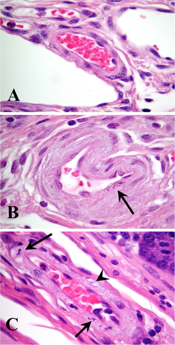
Figure 2.

Histological confirmation of vascular injury secondary to Shiga toxemia in ilea (submucosa). Arterioles in control pigs inoculated with non-pathogenic Escherichia coli lacked lesions (A). Arteriole from pig inoculated with Shiga toxin producing E. coli (STEC) strain S1191 and treated with the sham probiotic strain, which lacked the receptor Shiga toxin mimic (B). Segmental changes within the tunica media characterized by the presence of karyorrhectic nuclear debris (arrow) from a myocyte. Arteriole from pig inoculated with STEC strain S1191 and treated with the receptor mimic probiotic (C). Note nuclear remnants (arrows) of myocytes within the tunica media, as well as vacuolization (arrowhead) of the sarcoplasm. Hematoxylin and eosin, ×1000 magnification.
