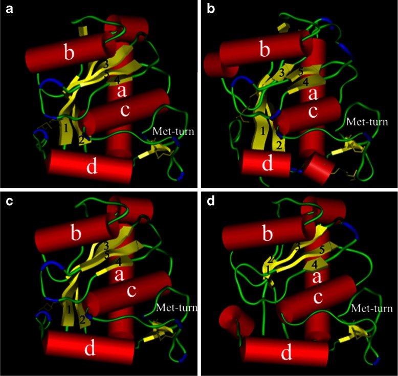
Fig. 3.
Molecular models of the four groups of C. s. scutulatus metalloproteinases. The three-dimensional molecular models of the four putative Crotalus s. scutulatus rattlesnakes metalloproteinases (quadrants I, II, III, and IV show models for GP1, GP2, GP3, and GP4 Mojave metalloproteinase models, respectively) as rendered in Kabsch–Sander mode. The a-helices have been rendered as red cylinders while the β-sheets illustrated as yellow flattened elongated arrows. The β-turns are rendered in blue and the disulfide bonds are illustrated as thick yellow lines bridging several regions of the enzyme

