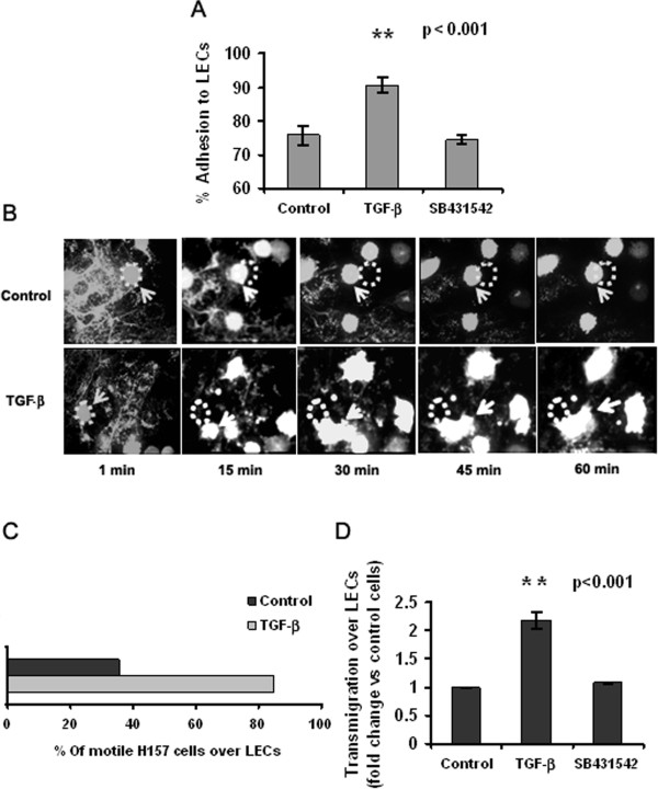
Figure 1.

TGF-β exposure enhances NSCLC cell adhesion and transmigration across lymphatic endothelial cells. (A) Adhesion of H157 cells to LEC monolayers after treatment for 5 days with TGF-β (2 ng/ml), and in the presence or absence of the TGF-βRI inhibitor SB 431542 (**p < 0.001, Student’s t-test). (B) Time-lapse microphotographs of the movement of H157 NSCLC cells, treated as in A, across LEC monolayers (63× water objective). Dots indicate the initial position of the cell and arrows indicate the same cell in motion. (C) Quantification of the number of H157 cells in transit over LEC monolayers, expressed as a percentage of the total number of cells counted in a single XY plane. (D) Quantification of NSCLC cell transmigration across monolayers of primary human LECs in the presence or absence of TGF-β. Data are presented as fold-change with respect to untreated control H157 cells (**p < 0.001, Student’s t-test).
