Figure 2.

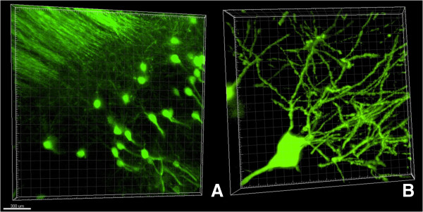
Confocal images of hippocampal neurons from line M Thy1-GFP mouse. (A) Neuronal structures of hippocampus, 20x objective. Scale bar is 300 microns. (B) 3D reconstruction of individual GFP-positive neuron, 60x objective.

Confocal images of hippocampal neurons from line M Thy1-GFP mouse. (A) Neuronal structures of hippocampus, 20x objective. Scale bar is 300 microns. (B) 3D reconstruction of individual GFP-positive neuron, 60x objective.