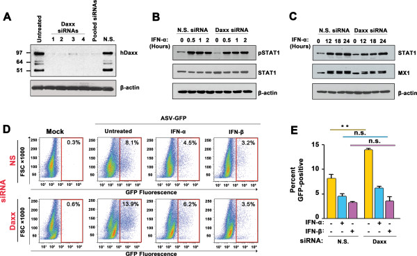
Figure 4.

Daxx is not required for type IFN inhibition of ASV replication in mammalian cells. (A) Immunoblot showing Daxx protein levels in HeLa cells following transfection of four individual (lanes 2–5) or pooled (lane 6) siRNAs to human Daxx. Lane 1 = untreated control. Molecular weight markers, in kilodaltons, are shown to the left. β-actin was used as a loading control. (B) Immunoblot analysis of STAT1 phosphorylation following IFN-α treatment of HeLa cells transfected with nonspecific (N.S.) or pooled Daxx siRNAs. Cells were treated with IFN-α (1000 U/ml) for the indicated time points. β-actin was used as a loading control. (C) Immunoblot analysis of STAT1 and MX1 induction following IFN-α treatment of HeLa cells transfected with nonspecific (N.S.) or pooled Daxx siRNAs. (D) ASV-GFP replication (indicated by % GFP-positive cells) in untreated, human IFN-α (1000 U/ml)- or human IFN-β (1000 U/ml)-treated HeLa cells 48 h post-infection. Cells were transfected with nonspecific (NS) or Daxx siRNAs for 72 h prior to infection, as indicted to the left. FACS data from are representative experiment are shown. FSC = Forward scatter. (E) Quantification of GFP-positive cells from three independent replicates of the experiment described in panel D. Error bars represent mean +/- standard deviation. **p <0.01. n.s., not significant.
