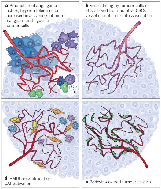
Figure 3. Potential mechanisms of resistance to targeted VEGF therapy in cancer.
Different mechanisms underlie the resistance to VEGF blockade seen in some patients with cancer. These mechanisms are not exclusive, and it is likely that several occur simultaneously in a single tumour. a, In established tumours, VEGF blockade aggravates hypoxia, which upregulates the production of other angiogenic factors or increases tumour cell invasiveness. Tumour cells that have acquired other mutations can also become hypoxia tolerant. The more malignant tumour cells are shown as dark green, blue and purple cells. b, Other modes of tumour vascularization, including intussusception, vasculogenic mimicry, differentiation of putative cancer stem cells (CSCs) into endothelial cells (ECs), vasculogenic vessel growth and vessel co-option (all denoted by the mosaic red–purple vessels), may be less sensitive to VEGF blockade. c, Tumour vessels covered by pericytes (green) are less sensitive to VEGF blockade. d, Recruited pro-angiogenic BMDCs (yellow), macrophages (blue and purple) or activated cancer-associated fibroblasts (CAFs; orange) can rescue tumour vascularization by the production of pro-angiogenic factors.

