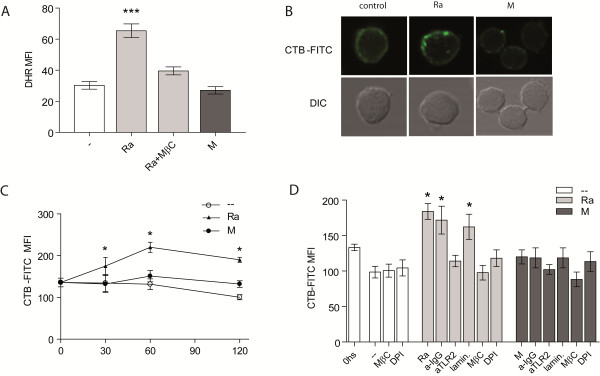
Figure 4.

Ra strain induces GM1 expression in PMN surface. (A) PMN treated with DHR were incubated with the lipid rafts inhibitor MβC, before challenge with 1:2 Mtb:PMN. Thereafter, the emission of oxidized DHR was evaluated by flow cytometry. Results are expressed as mean ± SE (n = 12); Ra vs. control (−−), Ra + MβC or M: ***p < 0.001 (B) PMN were cultured with bacteria and incubated with CTB-FITC and thereafter, lipid rafts were determined by the presence of clusters of GM1 expression by confocal microscopy. Propidium yodide (250 μg/ml) was used to visualize the nucleus. One of 4 experiments is depicted (C) PMN were cultured with a 2:1 Mtb:PMN ratio for 30, 60 o 120 min. Thereafter, cells were labeled with CTB-FITC (5 μg/ml) and fluorescence evaluated by flow cytometry. Results are expressed as mean ± SE (n = 7); control (−−) vs. Ra: *p < 0.05. (D) PMN were incubated with the ROS inhibitor DPI, MβC, laminarin, anti-TLR2 or irrelevant antibody for 30 min. at 37°C before challenge with Ra or M at 2:1 Mtb:PMN ratio. Thereafter, cells were labeled as described in (C). Results are expressed as mean ± SE (n = 7); control (−−) vs. Ra, laminarin or irrelevant: *p < 0.05.
