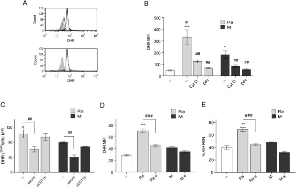
Figure 6.

α-glucans and non-opsonized phagocytosis are involved in ROS production. (A) PMN were pretreated with DHR and incubated with 1:2 Mtb:PMN ratio for 90 min (black line), pre-incubated with cytochalasin D (gray line) or, PMN without treatment (filled gray). Alternatively, PMN were pre-treated with anti-CD11b, second histogram (gray line). Representative out of 6 experiments are depicted (B) PMN pretreated with DHR were incubated with cytochalasin D or DPI prior to incubation with 10:1 Mtb:PMN ratio for 90 min. Results are expressed as a mean ± SE (n = 20); control (−−) vs. Ra ***p < 0.001; control (−−) vs. M *p < 0.05; Ra vs. M φp < 0.01; Ra vs. Ra + cytD or Ra + DPI: ##p < 0.01; M vs. M + cytD or M + DPI ##p < 0.01 (C) DHR-labeled Mtb (DHRMtb) were incubated with 5 × 10⁵ PMN at a 10:1 ratio for 90 min. Results are expressed as a mean ± SE (n = 20). Ra vs. Ra + serum ##p < 0.01; M vs. M + serum ##p < 0.01; Ra vs. M φp < 0.01 (D) PMN were pretreated with DHR and incubated with 1:2 Mtb:PMN ratio for 90 min. The α-glucans of clinical strains were eliminated (Ra e, M e) or not (Ra, M) as described in Methods. Results are expressed as a mean ± SE (n = 15); control (−−) vs. Ra ***p < 0.001; Ra vs. Ra e: ###p < 0.001. In all cases emission of oxidized DHR was evaluated by flow cytometry (E) Apoptosis was measured in 18-hr cultured PMN in the same conditions described in (A). Results are expressed as percentage of AV-FITC positive cells (% PMN AV + ± SE); control (−−) vs. Ra **p < 0.01; Ra vs. Ra e: ###p < 0.001.
