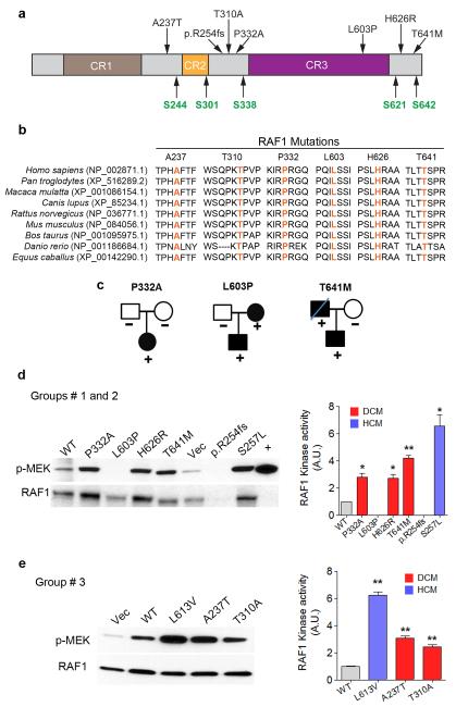
Figure 1. RAF1 mutants observed in dilated cardiomyopathy.
a. Schematic representation of RAF1 structure and location of residues altered in DCM patients. CR1-CR3 represents conserved regions of RAF1. Regulatory serine residues are shown in green. b. Alignment of RAF1 protein sequences from different species with the amino acid residues altered in DCM shown in orange. c. Pedigrees of DCM families with their RAF1 amino acid change indicated. d and e. RAF1 kinase assays. Vector alone (Vec), full-length wild-type (WT), HCM-associated (p.Leu613Val and p.Ser257Leu), and DCM-associated (p.Ala237Thr, p.Thr310Ala, p.Pro332Ala, p.Leu603Pro, p.His626Arg, p.Thr641Met and p.R254fs) RAF1 proteins were expressed in HEK293 cells as indicated. RAF1 was immunoprecipitated from EGF-stimulated cells at 15 min along with a positive control (+). Linked kinase assays were performed using inactive MEK1. RAF1 and phosphorylated MEK1 (p-MEK) were detected with anti-RAF1 (lower row) and anti-p-MEK (upper row) antibodies. Activation (p-MEK/RAF1) is expressed as Relative Expression compared to level in the WT cells. Data are mean values ± SD of two independent experiments. *p <0.05 or **p <0.01 vs. WT.

