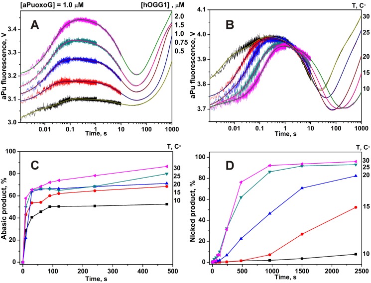
Figure 4. Stopped-flow fluorescence traces for interactions of hOGG1 with oxoG-substrate.
Changes in aPu fluorescence intensity during interaction of hOGG1 with oxoG-substrate at different concentrations of enzyme at 25°C (A) and at different temperatures (B). Solid lines represent the fitted curves. The kinetics of the accumulation of the abasic (C) and nicked (D) products, formed in the N-glycosylase and AP-lyase reactions, respectively, as detected in the PAGE experiments. The concentrations of hOGG1 and DNA for (B), (C) and (D) panels were 2.0 µM and 1.0 µM, respectively.

