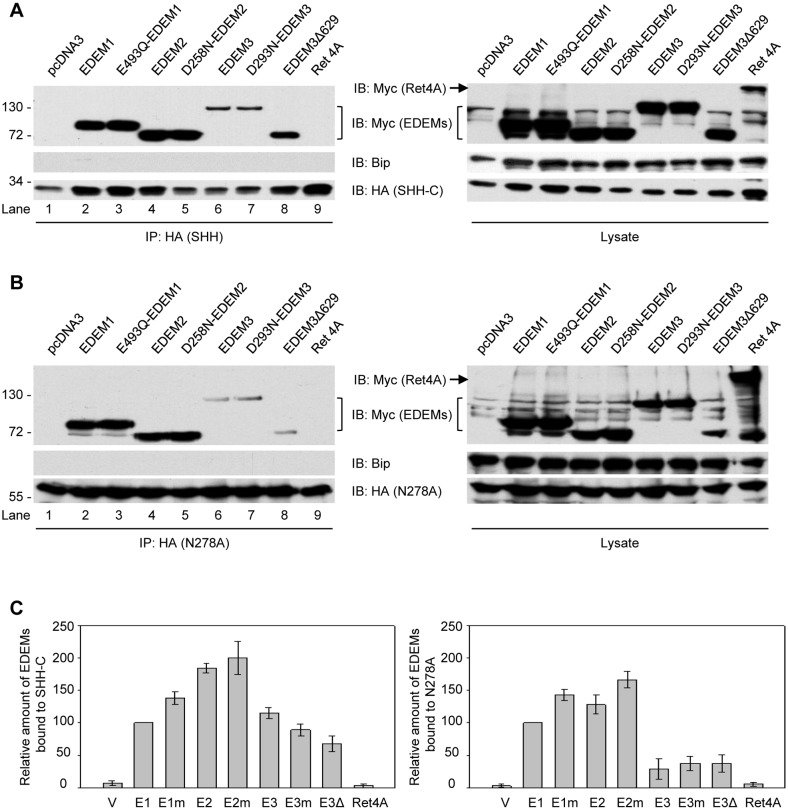
Figure 5. Interaction of SHH proteins with EDEMs.
(A) Cells were lysed in buffer containing 1% Triton and immunoprecipitated by HA antibodies. The precipitates and total cell lysate were separated in SDS-PAGE and immunoblotting with HA and Myc antibodies. As control, empty plasmid vector or plasmid encoding ER membrane protein Ret4A was transfected. (B) As in (A), but with N278A-HA plasmids. (C) Quantification of the amount of EDEMs bound to SHH-C (left) or N278A (right). Error bars were standard deviations from three independent repeats.

