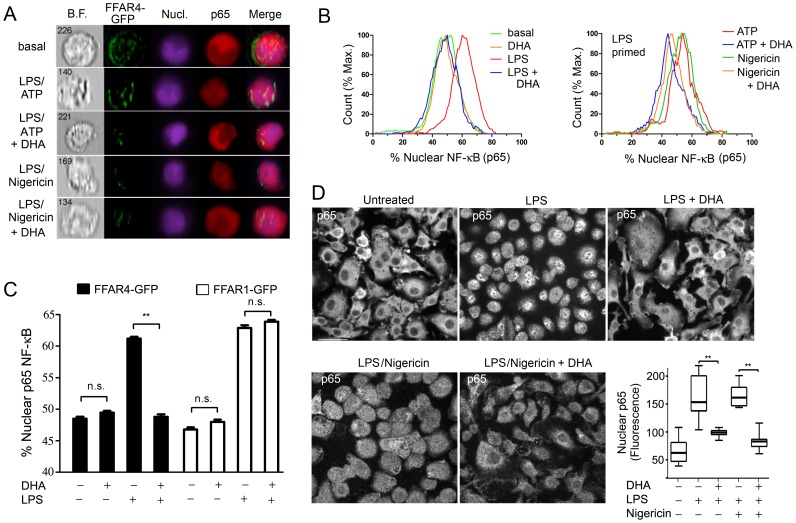
Figure 2. DHA inhibits the translocation of NF-κB to the nucleus.
FFAR4-GFP expressing LPS primed non-differentiated THP-1 cells were stimulated with ATP or nigericin. DHA (100 µM) was added or not at the priming step. PFA fixed cells were permeablized and then stained with p65 antibody and analysis was performed using an ImageStream instrument. Shown are the (A) Imaging results and (B) Flow cytometry results. (C) ImageStream analysis of nuclear p65 NF-κB in non-differentiated THP-1 cells expressing FFAR1-GFP or FFAR4-GFP and exposed to LPS, or not, in the presence of DHA (50 µM), or not. (D) Confocal microscopy of BMDMs LPS primed and treated with nigericin in the presence, or absence of DHA, to examine the status of NF-κB translocation by p65 immunostaining. Shown are representative individual images. Scale bar is 20 µM. Whisker plot shows the amount of nuclear p65 immunofluorescence in the nuclei of BMDMs treated as indicated. Data are representative of three independent experiments. **P<0.001, n.s-not significant.

