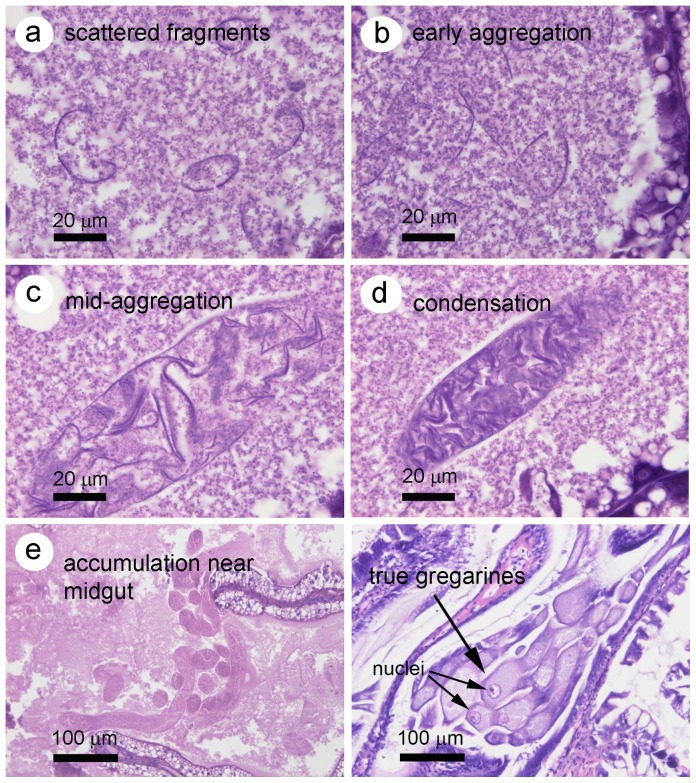
Figure 4. ATM aggregation steps in H&E stained HP tissue sections in comparison to true gregarines.
(a) Small, scattered membrane-lie structures in the HP tubule lumen. (b) More extended membranes beginning to aggregate in the tubule lumen. (c) Tighter aggregation of membranes bound by a continuous outer membrane and taking the shape of ATM. (d) Highly condensed ATM in a tubule lumen. (e) Accumulation of many individual ATM at the center of the HP near the midgut junction. (f) True gregarines clustered near the midgut junction and showing prominent nuclei.

