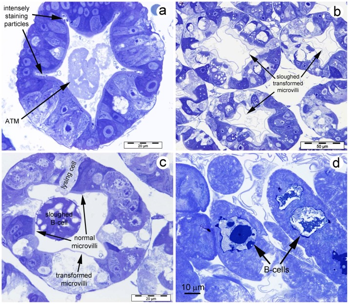
Figure 5. Semi-thin sections of HP tissue stained with toluidine blue.
(a) Cross section of an HP tubule near the distal end showing densely stained particles in crypts formed by folds of the tubule epihelium and showing aggregated, transformed microvilli (ATM) in the tubule lumen. Note that microvillar layers of all the cells are intact. (b) Cross sections of HP tubules showing sloughed, transformed microvilli. (c) Cross section of an HP tubule showing a modified, sloughed B-cell in the tubule lumen with microvilli scattered over its surface. Also seen are tubule epithelial cells with normal microvilli and transformed mivrovilli, and one cell denuded of microvilli, undergoing lysis. (d) High magnification of clustered ATM at the center of the HP clearly showing an outer membrane enclosing multitudes of folded transformed microvilli. Some also contain enclosed, sloughed B-cells. Note many free transformed microvilli fragments surrounding the ATM.

