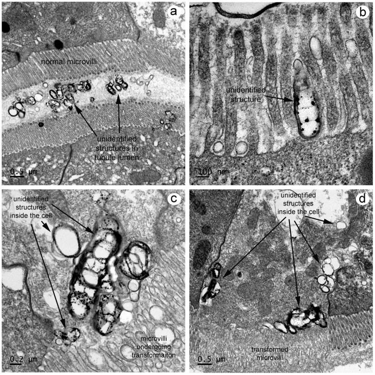
Figure 8. TEM of unusual electron-dense particles in HP tubule crypts.
(a) Low magnification of electron-dense particles of highly variable shape in the HP tubule lumen between layers of normal microvilli from facing epithelial cells. (b) High magnification of one of the electron-dense particles between the microvilli on the outside surface of an epithelial cell, possibly prior to cell entry. (c) High magnification of electron dense particles inside an epithelial cell with adjacent microvilli on the cell surface undergoing morphological changes. (d) Low magnification of an epithelial cell containing large numbers of electron dense particles and with microvilli in an advanced stage of transformation.

