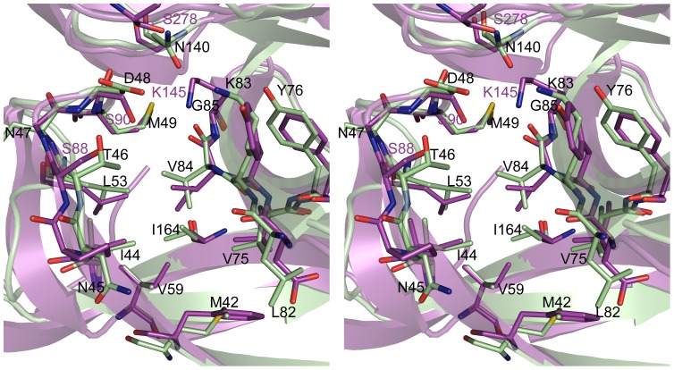
Figure 4. Residues lining SipA and E. coli SPase-I substrate binding pockets.
Stereo-view of residues lining the substrate-binding pocket of SipA (green) and SPase-I (magenta). SipA residues (one letter code) are labeled in black text, with the SPase-I catalytic residues labeled in magenta. The pockets have the same orientation as Figures 1 and 2 .

