Figure 1.
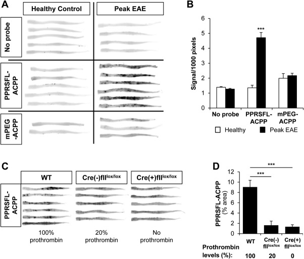
Specific detection of thrombin activity in the experimental autoimmune encephalomyelitis (EAE) spinal cord. (A) Whole spinal cord scans at 700nm from mice at peak EAE or healthy controls, injected with Cy5-labeled thrombin-specific PPRSFL–activatable cell-penetrating peptide (ACPP) or Cy5-labeled noncleavable control methoxy poly (ethylene glycol) (mPEG)-ACPP show specific uptake (dark spots) of PPRSFL-ACPP, indicative of increased localized thrombin activity at the peak of EAE. Uninjected healthy control and EAE mice are also shown as controls (no probe). (B) Quantification of total fluorescent signal in whole spinal cord scans from A, corrected for size. Data are presented as mean ± standard error of the mean (SEM); ***p < 0.001, 2-way analysis of variance (ANOVA); n = 5 to 7 per group for no probe or PPRSFL-ACPP and 2 to 3 for mPEG-ACPP. (C) Genetic reduction or elimination of prothrombin abolishes localized thrombin activity detection in EAE. Whole spinal cord scans from 3 cohorts of mice injected with PPRSFL-ACPP and polyI:C at EAE peak: wild-type (WT; 100% prothrombin), Mx-1Cre(−)fIIlox/lox (20% prothrombin), and Mx-1Cre(+)fIIlox/lox (no prothrombin). Prior to Cre recombinase induction, homozygous Mx1-Cre:fIIlox/lox mice exhibit baseline circulating prothrombin levels that are ∼20% of normal, whereas intraperitoneal injection of poly-I:C over a 6-day period results in a rapid loss of hepatic prothrombin expression and a near-complete loss (<5%) of circulating prothrombin within 5 to 6 days. Poly-I:C was administered at the time of overt clinical disease onset. (D) Quantification of PPRSFL-ACPP signal in whole spinal cord scans from C shows significantly reduced PPRSFL-ACPP retention with lower thrombin levels. Data are presented as mean ± SEM; ***p < 0.0001, 1-way ANOVA; n = 5 to 6 per group.

