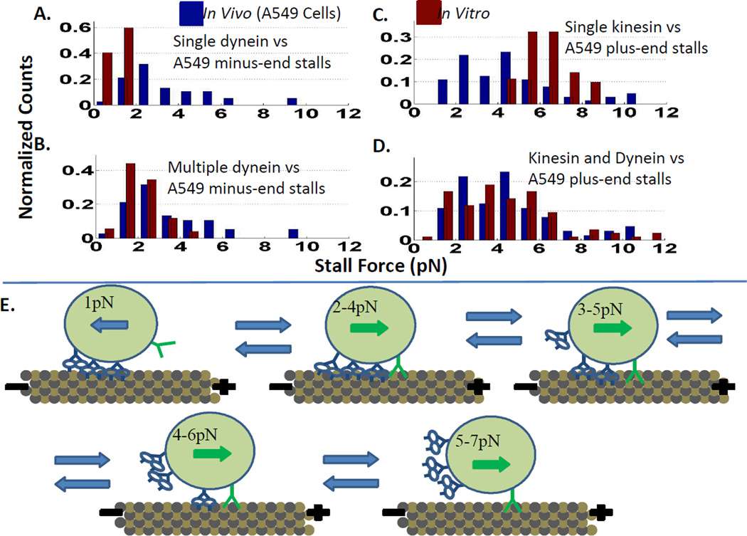
Figure 8.
Dynein drags. A. In vitro (red) beads with a single dynein have a lower stall force than minus-end directed lipid droplets in vivo (blue). B. Beads coated with multiple dynein in vitro have stall forces similar to minus-end directed lipid droplets in vivo. C. A single kinesin on a bead in vitro has a narrower and higher stall force distribution than plus-end directed lipid droplets in vivo. D. In vitro, beads coated with kinesin and dynein stall at similar forces while walking towards the microtubule plus-end as lipid droplets and phagosomes in vivo (less than the stall force of a single-kinesin)12a. E. Dynein being dragged by kinesin during plus-end directed motion appears to be the simplest explanation for why the beads with kinesin and dynein in vitro, and organelles in vivo, have reduced plus-end directed stall forces. Adapted with permssion from ref. 12a. Copyright 2013 National Academy of Sciences.

