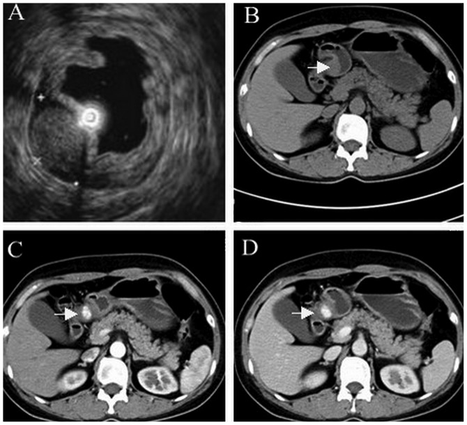
Figure 2.
(A) EUS an ovoid, demarcated, heterogeneous, hyperechoic tumor 18.6×11.8 mm in size, originating from the fourth EUS layer (muscularis propria). Abdominal computed tomography revealed a well-demarcated, ovoid mass at the antrum (arrow) on (B) unenhanced, (C) arterial- and (D) delayed-phase scans. EUS, endoscopic ultrasound.

