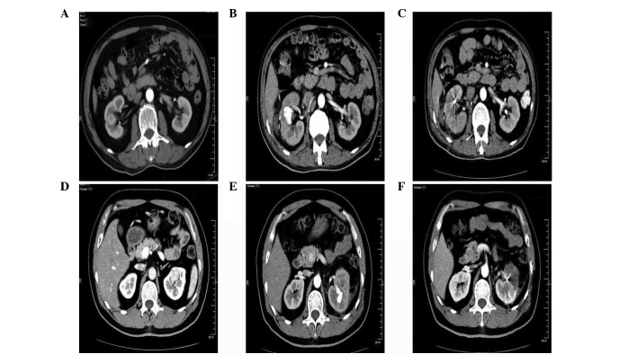
Figure 1.
Different situations of the lesions of the two cases through physical examination. Case one showed: (A) A 2.×2.5-cm space-occupying lesion in the center of the lower pole of the right kidney parenchyma; (B) a 6-cm hematoma on the edge of the right kidney and a 2.2-cm cystic shadow bound to the surgical area of right kidney with enhancement; and (C) superselective embolizations of the renal artery branches. Case two showed: (D) A 2.5×3.0-cm space-occupying lesion in the center of the upper pole of the left kidney parenchyma; (E) a ~3-cm cystic shadow bound to the center of the upper pole of the left kidney with enhancement; and (F) superselective embolizations of the renal artery branches.

