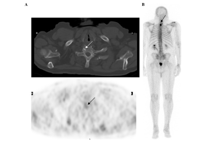
Figure 1.
(A) Transaxial fluorine-18 fluorodeoxyglucose (FDG) positron emission tomography (PET)-computed tomography (CT) images of the upper chest of patient 13. The 70-year-old male exhibited a prostate-specific antigen relapse (PSA level, 67 ng/ml) five years following radical prostatectomy. The first bone scintigraphy and pelvic CT were obtained one month prior to the FDG PET-CT and the two were negative. FDG PET-CT demonstrated a small sclerotic density with mild uptake (SUVmax, 3.5), which indicated metastasis in the right-side of the T1 vertebral body (indicated by the arrows). (B) Repeat bone scintigraphy one month later confirmed the FDG PET-CT-identified lesion (indicated by the arrow).

