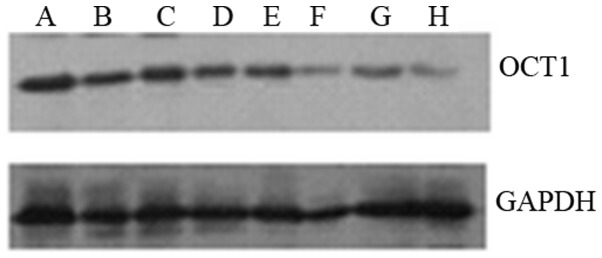
Figure 2.

Expression levels of the OCT1 protein in cervical cancer and the adjacent non-cancerous tissues. In total, (lanes A, C, E and G) four cervical cancer and (lanes B, D, F and H) four of the adjacent non-cancerous tissues were selected to detect the expression levels of OCT1 protein by western blot analysis. Data are representative of three independent experiments. OCT1, octamer transcription factor 1; GAPDH, glyceraldehyde-3-phosphate dehydrogenase.
