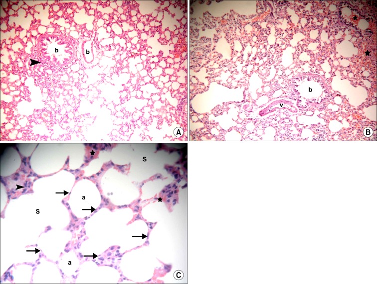
Fig. 3.
(A) Section in the lung of a rat in IVI stem cell therapy group showing two bronchioles (b), one of them demonstrates cellular infiltrate in the adventitia (arrowhead) and a congested vessel (c) (H&E, ×100). (B) Section in the lung of a rat in IVI stem cell therapy group showing a bronchiole (b), a vessel with partially thickened wall (v), congested vessels (c) and extravasated RBCs (*) (H&E, ×100). (C) Section in the lung of a rat in IVI stem cell therapy group showing normal alveoli (a) and alveolar sacs (S) lined by multiple cells exhibiting flat nuclei (arrows). Note cellular infiltrates (arrowheads) and extravasated RBCs in few alveolar septa (*) (H&E, ×400).

