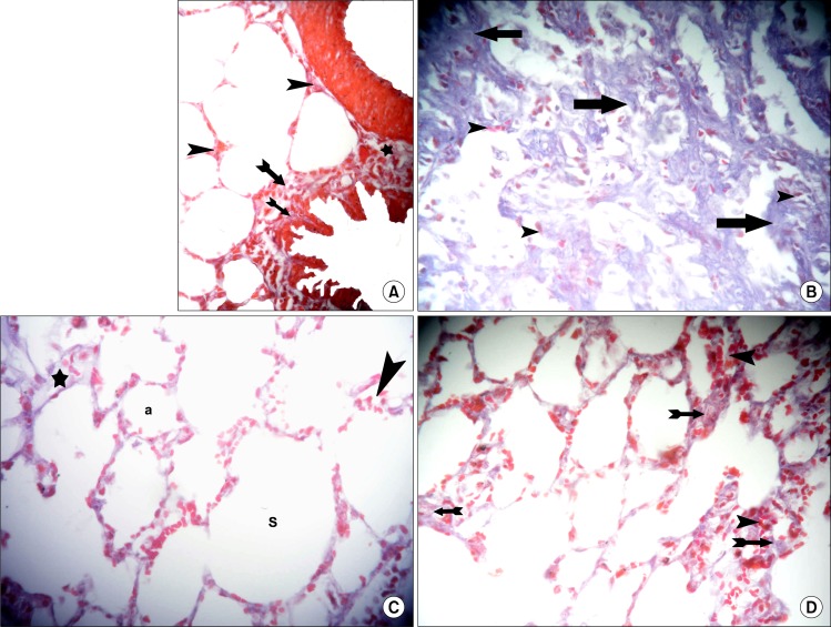
Fig. 5.
(A) Section in the lung of a control rat showing fine collagen fibers (arrowheads) in the alveolar septa, denser collagen fibers in the wall of a bronchiole (arrows) and in the adventitia (*) of a blood vessel (Masson’s trichrome, ×400). (B) Section in the lung of a rat in amiodarone group showing dense collagen fibers (arrows) and infiltrating cells (arrowheads) in the alveolar septa (Masson’s trichrome, ×400). (C) Section in the lung of a rat in IVI stem cell therapy group showing dense collagen fibers (*) in a septum and infiltrating cells (arrowhead) in another septum around normal alveoli (a) and alveolar sacs (S) (Masson’s trichrome, ×400). (D) Section in the lung of a rat in IPI stem cell therapy group showing dense collagen fibers (arrows) in some septa and infiltrating cells (arrowheads) in other septa (Masson’s trichrome, ×400).

