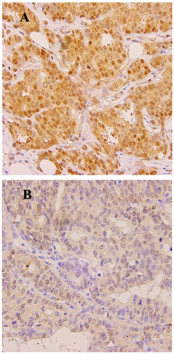
Figure 1.

Representative photomicrographs of immunohistochemical staining for MGMT in breast cancer (magnification, ×100). Positive staining is denoted by brown-stained nuclei and extremely weak cytoplasmic staining. (A) MGMT positivity (fraction of positive cells >10%). (B) MGMT negativity. MGMT, O6-methylguanine-DNA methyltransferase.
