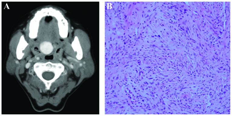
Figure 1.
Imaging and pathological findings of the solitary fibrous tumor in Case 1. (A) A well-circumscribed mass with homogenous enhancement measuring ~2 cm in diameter was detected in the soft palate on an enhanced computed tomography scan. (B) Hematoxylin and eosin-stained section of tumor showing storiform or fascicular pattern (original magnification, ×200).

