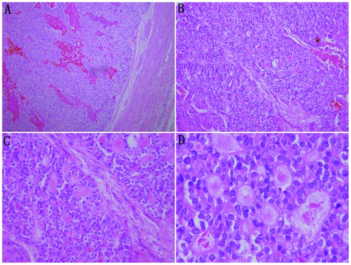
Figure 3.
Microscopic examination of the tumor in patient 1. (A) The tumor is encapsulated with a fibrous pseudo-capsule (×40). (B) The tumor cells are separated by mesenchymal tissues into patches with a morphology of ‘dried follicles’ (×100). (C) The tumor cells show a morphology mimicking thyroid follicles with red-stained colloid-like material in the lumen. The colloid-like material broke a number of the lumens and merged (×200). (D) The nuclei are enlarged and overlapped in round, oval or spindle shapes with fine chromatin pattern and one to two inconspicuous nucleoli per nucleus (×400). Hematoylin and eosin staining.

