
Figure 4. Quantitative analysis confirms decreased and delayed accrual of peak bone volume in fracture callus of HFD-fed mice.
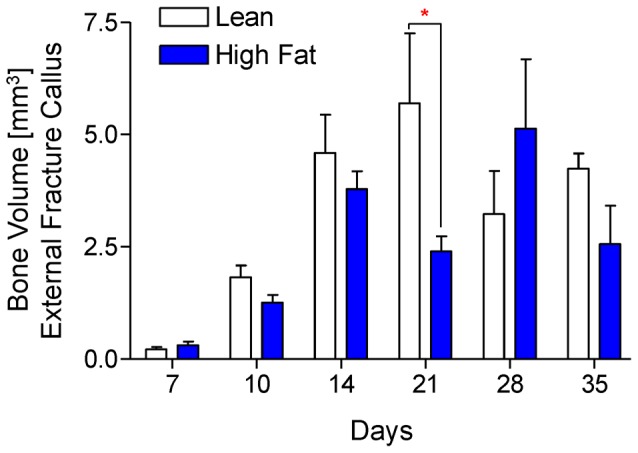
Using μCT scans that were used create the reconstructions shown Fig. 3, bone volume was quantified from lean- and HFD-fed mice at 7, 10, 14, 21, 28, and 35 days post-fracture. Bars represent means ± SEM (n≥5). *p<0.05 compared to time-matched lean diet controls using two-way, unpaired ANOVA with Bonferroni post-tests.