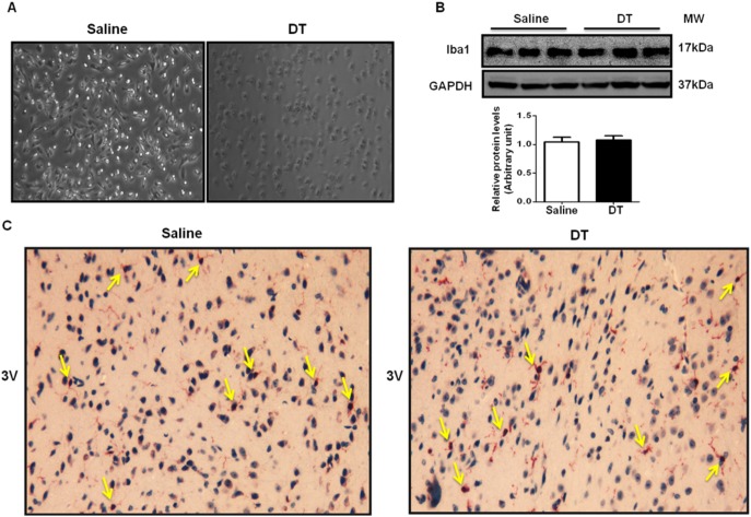
Figure 4. DT depletes microglia in.
Microglia were isolated from day 1 newborn LysMCre/iDTR mice (n = 3). Isolated microglia were cultured in cell-culture plates. Saline or DT (0.25 ng/ml) was treated for 8 hrs. (A) Representative macroscopic images of isolated microglia treated with saline or DT for 8 hrs. (B) Total protein levels of microglia marker Iba1 in the hypothalamus of mice treated with saline or DT for 6 days. (C) Microglia in the brain were identified by immunohistochemisty using microglia marker Iba1 antibody. Arrows indicate positive cells (n = 3). 3 V represents the direction of third ventricle.

