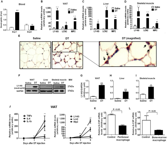
Figure 6. Macrophage depletion increases tissue NE infiltration.
Male LysMCre/iDTR mice were fed a chow diet and were i.p. injected with DT (10 ng/g) or saline. (A) Blood NE concentration was assessed after 1 and 6 days of DT injection by a hematology test. (B–D) mRNA levels of NE markers LY-6G, LCN, and NE-chemoattractant chemokine MIP2 in WAT, liver, and skeletal muscle were measured by Q-PCR (n = 5). LY-6G, lymphocyte antigen 6 complex, locus G; MIP2, macrophage inflammatory protein 2; LCN2, lipocalin 2 (E) Infiltrated NEs in WAT were identified by immunohistochemisty using NE marker LY-6G antibody. Arrows indicate positive cells. (F) Protein levels of NE marker LY-6G in WAT, liver, and skeletal muscle were measured by Western blotting (n = 4 each). Representative images were shown in the figure. (G–I) Quantification of LY-6G/GAPDH protein levels in tissues. (J) The comparison of inflammatory cytokine expression with NE marker expression in WAT by Q-PCR during the different time points of DT treatment. (K and L) G-CSF mRNA levels were measured by Q-PCR using RNA samples from OP9 cells treated with 10% peritoneal (K) or bone-marrow derived macrophage (L) conditioned media (n = 4 each). Data are represented as mean ± SEM. Asterisks denote significant differences *P<0.05, **P<0.01 vs. saline-treated mice.

