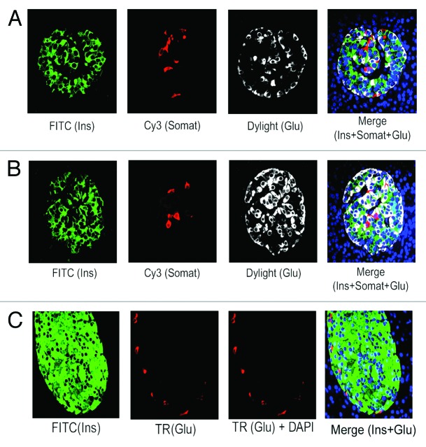
Figure 1. Prevalence of β-cells in adult human pancreatic islets. Analysis of the abundance of β-, α-, and δ- cells in normal adult pancreatic tissue sections, obtained from donors (age: 30–50 y) (A and B) and mice (age: 9 mo) (C), by immunoflluorescence and confocal microscopy. The tissue sections were incubated with the specific primary antibodies against insulin, somatostatin and glucagon followed by incubation with the appropriate secondary antibodies conjugated with either FITC (for insulin) or Cy3 (for somatostatin) or Dylight/Texas Red (TR) (for glucagon). For (A and B), insulin, somatostatin and glucagon are represented by green, red, and white, respectively and for (C), insulin and glucagon are indicated by green and red, respectively. For (A–C), DAPI-stained nuclei are represented by blue. Almost 200 individual islets (10–15 islets/section) for (A and B) were analyzed to assess the abundance of β-, α-, and δ-cells. (A) represents the distribution pattern of β-, α-, and δ-cells found in majority (80%) of adult human islets, whereas, (B) represents the distribution pattern of these cell types found only in 20% of adult human islets. (C) shows the typical distribution of β- and α-cells in adult mouse islets. In this and subsequent figures, insulin, somatostatin, and glucagon are abbreviated as Ins, Somat, and Glu.

An official website of the United States government
Here's how you know
Official websites use .gov
A
.gov website belongs to an official
government organization in the United States.
Secure .gov websites use HTTPS
A lock (
) or https:// means you've safely
connected to the .gov website. Share sensitive
information only on official, secure websites.
