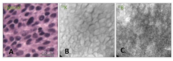
Fig.2.

SIMS imaging analysis of B16 tumor from cis-ABCPC treated B16 melanoma bearing mice. The optical image was recorded from H&E stained 4 μm thick tumor cryosection. SIMS images were recorded from adjacent 4 μm thick cryo-sections. The SIMS 39K+ image (B.) was integrated on the CCD camera for 0.2 sec and 11B+ image (C.) for 2 min.
