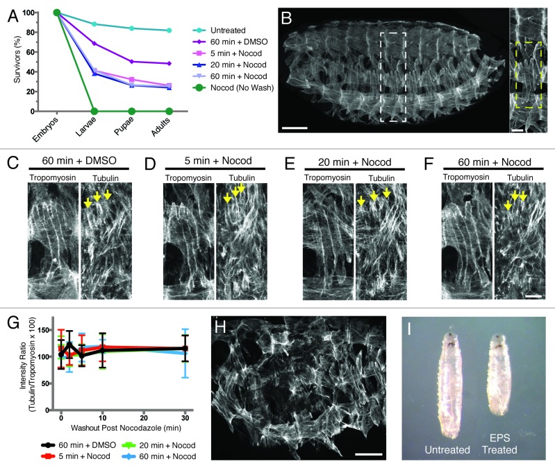
Figure 2. EPS is not sufficient to permeabilize late-stage embryos and is detrimental to normal Drosophila development. (A) Graph of viability of 1:5::EPS:PBS-treated embryos following washout of either drug or vehicle control (unless otherwise noted). (B, left) Wild-type stage 16 (16 h AEL) Drosophila embryo oriented with anterior to the left and dorsal up, immunostained for Tropomyosin to show muscles. Scale bar, 50 μm. (Right) Higher magnification of white dashed box outlining 1 hemisegment. Yellow dashed box highlights the 4 Lateral Transverse (LT) muscles within a hemisegment. Scale bar, 10 μm. (C–F) Stage 16 embryos treated for the indicated length of time with 1:5::EPS:PBS and either DMSO vehicle control or 66 nM Nocodazole in DMSO, removed from drug/EPS solution without further washout, immediately fixed, and immunostained with α-Tubulin and Tropomyosin antibodies to label the microtubules and the muscle cells, respectively. Arrows indicate selected microtubules near the dorsal poles of the LT muscles. Scale bar, 10 μm. (C) Sixty minute treatment with 1:5::EPS:PBS and DMSO vehicle control. (D) Five minute treatment with 1:5::EPS:PBS and 66 nM Nocodazole in DMSO. (E) Twenty minute treatment with 1:5::EPS:PBS and 66 nM Nocodazole in DMSO. (F) Sixty minute treatment with 1:5::EPS:PBS and 66 nM Nocodazole in DMSO. (G) Quantification of immunofluorescence intensity of α-Tubulin detected in each treatment condition shown in (C–F) over time after the removal of Nocodazole. (Note: all 4 data sets fall on top of one another.) (H) Stage 16 embryo treated with DMSO vehicle control in 1:5::EPS:PBS for 20 min immunostained for Tropomyosin (compare with (B). Scale bar, 50 μm. (I) Comparison of larval size in L3 wandering larvae.

An official website of the United States government
Here's how you know
Official websites use .gov
A
.gov website belongs to an official
government organization in the United States.
Secure .gov websites use HTTPS
A lock (
) or https:// means you've safely
connected to the .gov website. Share sensitive
information only on official, secure websites.
